Center for Genomically Engineered Organs
Fluorescent In Situ Sequencing (FISSEQ) of RNA molecules in cultured human fibroblasts
n this animated image, each frame represents a cycle of sequencing and each dot represents a single RNA molecule in a cell. The color of a dot indicates the RNA base identified during the cycle, so that the series of colors seen over the course of the animation reads off a small segment of the RNA's sequence. Because each cell's RNA profile can be seen separately, FISSEQ makes it possible to see differences in cell types and behaviors with cultured cells and complex tissue samples.
(Images courtesy of the Church lab.)
(Images courtesy of the Church lab.)
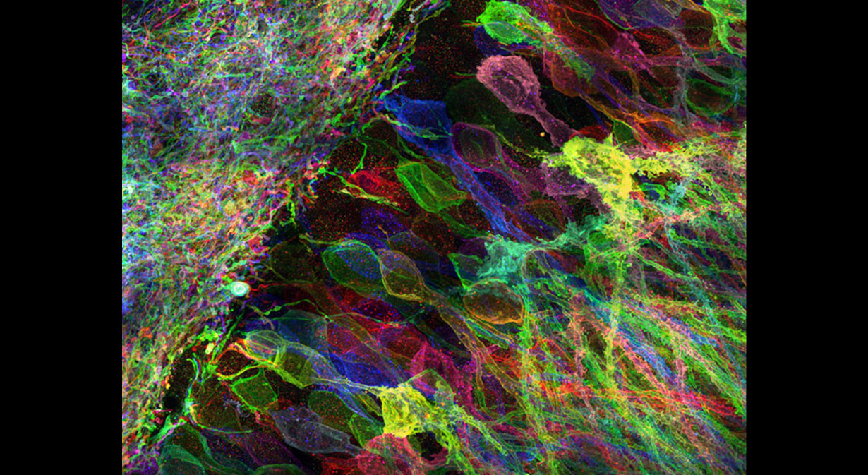
Mouse hippocampal neurons viewed with Expansion Microscopy
We are working to use the newly developed technique of Expansion Microscopy (ExM) to study our genomically engineered organoids at super-resolution. This image shows mouse hippocampal neurons, expressing Brainbow 3.0 delivered in viral form, that have been expanded ~4.5x linearly in each dimension after being processed with ExM.
(Image courtesy of the Boyden Lab.)
http://expansionmicroscopy.org/
(Image courtesy of the Boyden Lab.)
http://expansionmicroscopy.org/
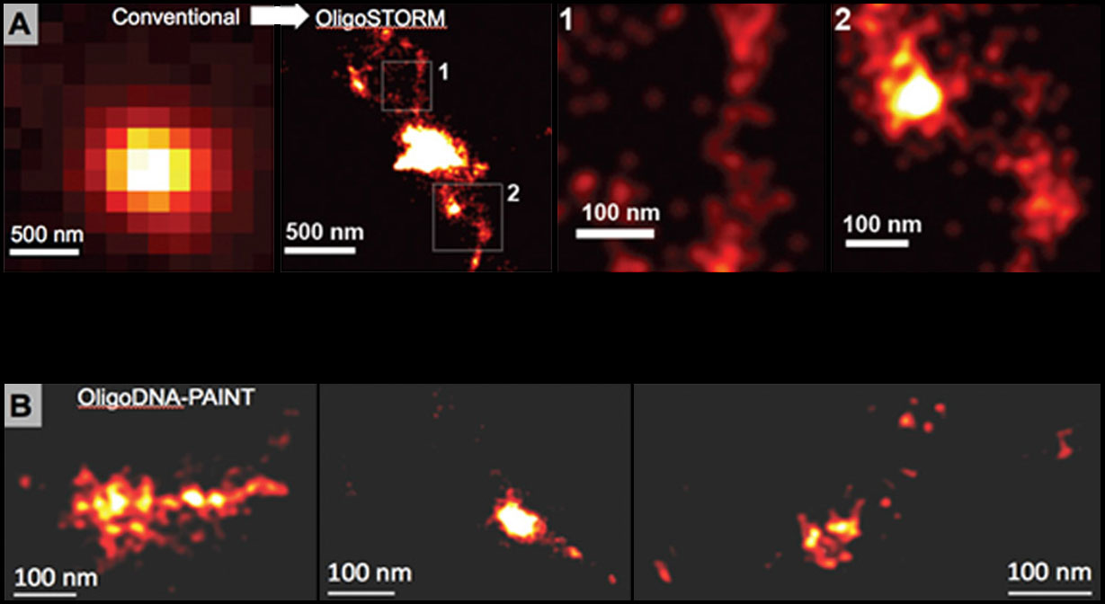
Super-resolution imaging of genomic loci labeled with OligoPaints
(A) ≤20 nm super-resolution OligoSTORM images of 2,394 oligos targeting 316 kb of the Drosophila BX-C complex. (B) OligoDNA-PAINT images of 106 oligos targeting 5 kb of the mouse hoxB locus.
(Images courtesy of the Wu, Zhuang, and Yin labs.)
(Images courtesy of the Wu, Zhuang, and Yin labs.)
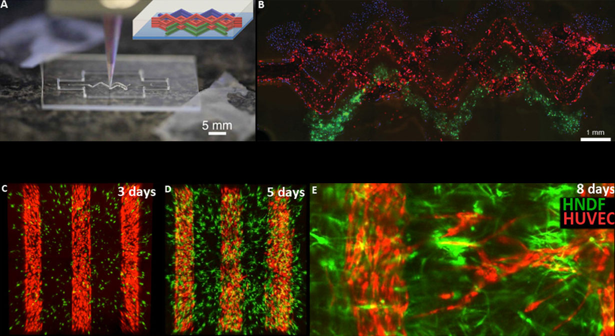
Cell-laden gel matrix encourages angiogenesis
To make organoids more like real organs, they must be vascularized. Vascular channels can be engineered into tissue cultures by 3D printing techniques that lay down channels that can be lined with human endothelial cells (HUVEC) within a gel matrix containing human fibroblasts (HDNF). Cell printing is shown on top: In A a print head is laying down one of three kinds of cells in a pattern (inset); B shows the final product. However, we want vessels to grow into the organoids and so are developing ways of leveraging the natural propensity of endothelial vessels to sprout new vessels, as shown at the bottom (C-E).
Copyright © The President and Fellows of Harvard College